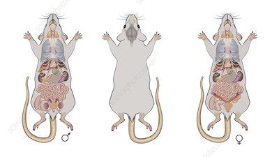
anatomic mouse|the anatomy of laboratory mouse : Tuguegarao Using genetic and viral labelling, barcoded anatomy resolved by sequencing, single-neuron reconstruction, whole-brain imaging and cloud-based neuroinformatics . Resultado da Mira los videos porno de MartinaOlvr y su perfil oficial, solo en Pornhub. Descubre los mejores videos, fotos, gifs y listas de reproducción de la .
0 · the anatomy of laboratory mouse
1 · mouse digestive system diagram
2 · mouse brain anatomy and function
3 · mouse anatomy pictures
4 · mouse anatomy pdf
5 · mouse anatomy map
6 · female reproductive mouse anatomy
7 · anatomy of a female mouse
8 · More
WEBConfira os diversos modelos do CapCut em raca do cachorro da lele burnier, incluindo cachorro de criatorioSalles, Cachorro de Motivacional [SV].
anatomic mouse*******Mouse - Anatomy atlas - CT: Digestive system, Urinary organs. Mouse - Pelvis - Anatomy: Urogenital system, Urinary bladder, Uterus. Cross-sectional anatomy of the mouse on high-resolution X .This textbook describes the basic neuroanatomy of the laboratory mouse. The reader will be guided through the anatomy of the mouse nervous system with the help of abundant .
The mouse remains the key animal model for exploring human disease and, despite its small comparative size, the laboratory mouse is anatomically similar to .
Using genetic and viral labelling, barcoded anatomy resolved by sequencing, single-neuron reconstruction, whole-brain imaging and cloud-based neuroinformatics .The laboratory mouse or lab mouse is a small mammal of the order Rodentia which is bred and used for scientific research or feeders for certain pets. Laboratory mice are usually of the species Mus musculus.
The mouse remains the key animal model for exploring human disease and, despite its small comparative size, the laboratory mouse is anatomically similar to .This paper presents a deformable mouse atlas of the laboratory mouse anatomy. This atlas is fully articulated and can be positioned into arbitrary body poses. The atlas can .
19. Cervical vertebra. 20. Thoracic vertebra. 21. 5th thoracic vertebra with rib. 22. Lumbar vertebra. 23. Sacrum. 24. Sacrum. As well as illustrating the normal microscopic anatomy of the mouse, the book also describes and explains the common anatomic variations, artefacts associated .Order your anatomy atlas from the AALAS Store!. Comparative Anatomy of the Mouse and Rat: a Color Atlas and Text provides detailed comparative anatomical information for those who work with mice and rats in animal research. Information is provided about the anatomical features and landmarks for conducting a physical examination, collecting .
12.4 Anatomy and histology of the eye and associated glands 199 12.5 Background and development of the ear 204 12.6 Sampling technique for the ear and associated structures 205 12.7 Anatomy and histology of the ear and associated glands 206 References 209 Chapter 13 Musculoskeletal system 211 Cheryl L. Scudamore 13.1 Background and . Did you know mouse organs are very similar to human organs? Learn how to dissect a mouse in this video, which also covers its external and internal anatomy a.
As well as illustrating the normal microscopic anatomy of the mouse, the book also describes and explains the common anatomic variations, artefacts associated with tissue collection and background lesions to help the scientist to distinguish these changes from experimentally- induced lesions. This will be an essential bench-side .Alibaba.comThe Anatomy of the Laboratory Mouse Margaret J. Cook CONTENTS Contents: Full Title Page Foreword Introduction Externals Skeleton Viscera Circulatory Systemanatomic mouse the anatomy of laboratory mouseThe Anatomy of the Laboratory Mouse Margaret J. Cook INDEX (The numbers refer to the illustrations. Those in italic denote Figures in which the subject is more fully treated.). Links to MGI go to the relevant term in the Adult Mouse Anatomy browser. Acetabular notch, 37 Acetabulum, 37, 38, MGI Acromion process, 31 Adrenal (suprarenal) gland, 60, 111, 112, . How does a mammal breath? How its reproductive system is done? «Mouse dissection» is a scientific movie and a pedagogic document for Biology students. The HD.19. Cervical vertebra. 20. Thoracic vertebra. 21. 5th thoracic vertebra with rib. 22. Lumbar vertebra. 23. Sacrum. 24. Sacrum.
anatomic mouseAmbesonne Human Anatomy Mouse Pad, Chart of Organs Body Structures Biology Themed Cartoon Schemes, Rectangle Non-Slip Rubber Mousepad, Standard Size, White and Multicolor. 4.4 out of 5 stars 346. $17.99 $ 17. 99. FREE delivery Sep 1 - 5 . Options: 4 sizes. Small Business. Small Business.Last but not least, this atlas includes more detailed anatomical structures than the existing mouse atlases, such as individual vertebrae, spinal cord, and neck brown fat, and therefore provides more anatomical details for atlas-based simulation. For the first time, we use multiple training subjects to construct a whole-body-scale mouse atlas.
Anatomy of a Monitor; Anatomy of a Mouse; Anatomy of a Keyboard; Anatomy of a Gamepad; We'll start by digging into the guts of a simple mouse – the kind that costs less than $10 and is used by . The Allen brain atlas. The Allen Mouse Brain Atlas (ABA, www.allenbrainatlas.org; Lein et al. 2007), a genome-wide atlas with datasets showing the expression about 21,000 transcripts (Ng et al. 2007) in the adult mouse brain (56d, C57BL/6 J; Allen Institute for Brain Science 2012).The distributions of most of these .
MOp-ul borders and cell types. The spatial location of rodent primary motor cortex (MOp) has been defined by cytoarchitecture, micro- or optogenetic- stimulation 28 and anatomical tracing 29, 30 .
Last but not least, this atlas includes more detailed anatomical structures than the existing mouse atlases, such as individual vertebrae, spinal cord, and neck brown fat, and therefore provides more anatomical details for atlas-based simulation. For the first time, we use multiple training subjects to construct a whole-body-scale mouse atlas.the anatomy of laboratory mouse Anatomy of a Monitor; Anatomy of a Mouse; Anatomy of a Keyboard; Anatomy of a Gamepad; We'll start by digging into the guts of a simple mouse – the kind that costs less than $10 and is used by . The Allen brain atlas. The Allen Mouse Brain Atlas (ABA, www.allenbrainatlas.org; Lein et al. 2007), a genome-wide atlas with datasets showing the expression about 21,000 transcripts (Ng et al. 2007) in the adult mouse brain (56d, C57BL/6 J; Allen Institute for Brain Science 2012).The distributions of most of these . MOp-ul borders and cell types. The spatial location of rodent primary motor cortex (MOp) has been defined by cytoarchitecture, micro- or optogenetic- stimulation 28 and anatomical tracing 29, 30 .

The mouse is anesthetized and then restrained manu-ally on a solid surface. The site of injection is approxi-mately half way between the eye and ear and just off the midline (Figure 32.16a). The recommended maxi-mum volume per suckling mouse is 0.01 ml and that. Figure 32.15 Intradermal injection into the back skin.

The mouse is anesthetized and then restrained manu-ally on a solid surface. The site of injection is approxi-mately half way between the eye and ear and just off the midline (Figure 32.16a). The recommended maxi-mum volume per suckling mouse is 0.01 ml and that. Figure 32.15 Intradermal injection into the back skin.The Laboratory Mouse, Second Edition is a comprehensive book written by international experts. With inclusions of the newly revised European standards on laboratory animals, this will be the most current, global authority on the care of mice in laboratory research. This well-illustrated edition offers new and updated chapters including .
This increased resolution enriches and expands existing human and mouse anatomic ontologies in multiple ways by 1) incorporating existing terms from non-anatomic ontologies into a well-defined anatomic ontology with hierarchical organizations and relationships and 2) incorporating additional terms that describe anatomic structures, . An anatomy ontology for mouse development: early versions. The anatomical ontology for the developing mouse originated as a Tissue Index for the second edition of The Atlas of Mouse Development (Kaufman 1994).The initial list comprised terms representing structures identified in serial histological sections of mice throughout the . Baylor College of Medicine (BCM) In a huge collaborative effort, millions of cells in the mouse brain have been mapped in detail. Two scientists examine the resulting wealth of insights into gene . Mouse Human; Nasal passage: Complex anatomy with olfaction as primary function; obligate nose breathers: Relatively simple anatomic structure with breathing as primary function; both nasal and oral breathing possible: Nasal vestibule: Surface is lined by squamous epithelium but lacks hair follicles:High resolution anatomical reference atlases and histology for mouse and human. View Atlases. Explore the Mouse Atlas/CCF. Common Coordinate Framework (CCFv3) is a 3D reference space by creating an average brain at 10um voxel resolution. Explore. Programmatic Access. Use the API and Python SDK to access reference data .
Anatomy of adipose depots. (A, A’) In mouse skin, dermal WAT (dWAT) forms a continuous layer (shown in yellow) separated from subcutaneous WAT (sWAT) by the panniculus carnosus muscle (shown in green). This separation is not prominent in human skin, where dWAT is continuous with underlying sWAT (orange) (A’). Dermal .
lyrics aud-20221014-wa0002 : A plataforma mais confiável lyrics aud-20221014-wa0002 Newry City. Assistir Agora. Futmax é a plataforma perfeita para assistir futebol ao vivo de qualidade, livre dos .
anatomic mouse|the anatomy of laboratory mouse